توده های فک و صورتCT/RADIOLOGY NECK AND HEAD
تصویربرداری سه بعدی ۳D IMAGING
BY: DR AMIRAFRAZ FALAH, DR SOFIA SABOURI
:Patient history
Middle age gentleman presented with sensation of bulging in left side of lower jaw without any
history of recent surgery
Imaging findings (CT scan)
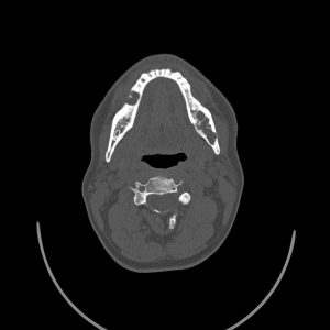
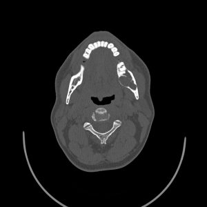
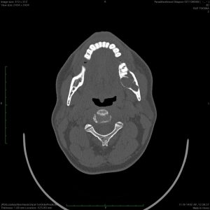
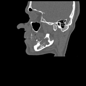
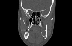
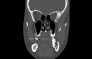
There is a lytic expansile lesion measuring 17*21mm in left side of mandibular body adjacent to root
of last left molar tooth. Thinning of bony margins and some erosions in superior margin of this
lesion is detected. Also, some erosions adjacent to root of aforementioned molar tooth are present.
Some septations and loculations are evident into this lesion. This lesion is located in close vicinity
with medial site of mandibular canal and shows abutment with inferior alveolar nerve. No evidence
of extension to surrounding soft tissues is present. This lesion is most probably in favor of an an
.ameloblastoma. However odontogenic keratosis however is considered in DDX less likely
:Diagnosis: (with pathology confirmation)
Ameloblatoma
:Ameloblastoma
Ameloblastomas are locally aggressive benign tumors that arise from the mandible, or less
commonly, from the maxilla. They typically occur as hard, painless lesions near the angle of the
mandible in the region of the 3rd molar tooth. it is a locally aggressive neoplasm with a high rate of
recurrence. Approximately 20% of cases are associated with dentigerous cysts and unerupted teeth
Multicystic ameloblastomas account for 80-90% of cases which are classically expansile “soapbubble” lesions, with well-demarcated borders and no matrix calcification. Resorption of adjacent
teeth and “root blunting” is often a feature. When larger it may also erode through the cortex into
.adjacent soft tissues
Unicystic ameloblastomas are well-demarcated unilocular lesions that are often pericoronal in
position. These are commonly found in the posterior mandible, particularly at the molars. They are
indistinguishable from other unilocular pericoronal lesions, such as dentigerous cysts, ameloblastic
.fibromas and odontogenic keratocysts on CT
Differential diagnosis:
۱- dentigerous cyst
۲- odontogenic keratocyst (OKC)
۳- aneurysmal bone cyst (ABC)
۴- fibrous dysplasia
