
Tooska interesting cases
Case 1
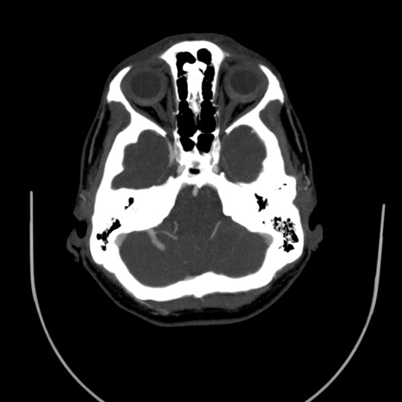
A 36 year old female with recent headache and nausea-vomiting since 2 weeks ago. No specific history of CVA or neurological disease. A suspicious vascular lesion was reported in the Brain MRI and referred for brain CT angiography.
Radiologic findings
A vascular structure as a network which drains to one vessel. It shows multiple branching appearance in right cerebellar hemisphere (Caput Medusa Sign). Finally the vessel drains to right transverse sinus. No evidence of obvious connection to arterial vessels. No evidence of surrounding hemorrhage.
Differential diagnosis
Deep venous anomaly (cerebral venous angioma) — congenital malformation, the most common cerebral vascular malformation. Characterised by multiple veins with appearance of caput medusa or palm tree, draining into a single larger vein which finally drains into a dural sinus or an ependymal vein.
Two common types: frontoparietal region (36–64%) draining towards the frontal horn of the lateral ventricle, and cerebellar hemisphere (14–27%) draining towards the 4th ventricle.
Other differentials: arteriovenous malformation, dural sinus thrombosis or dural arteriovenous fistula with collateral transparenchymal drainage, Sturge-Weber syndrome with leptomeningeal angiomatosis, and demyelination (which may also have enlarged medullary veins).
Associations of DVA
Usually solitary (75%). About 20% (range 8–33%) of cases are associated with cavernous malformations, venous malformations of the head and neck, and (uncommonly) cortical dysplasia.
Case 2
A 60 year old man referring for chest CT scan with long term history of dyspnea and cough. Symptoms have been exacerbated since 6 months ago. Normal echocardiography and exercise test.
Radiological findings
Few honeycombing, diffuse tractional bronchiectasis and irregular interlobular septal thickening in periphery of both lungs. Almost complete collapse of LLL, RML and anterior segment of RUL associated with significant narrowing of their airways, with some calcified foci in the aforementioned collapses. Small patches of consolidation and ground glass opacities in apices of both lungs.
Scattered centrilobular nodules in both lungs. Bilateral hilar lymphadenopathy containing calcifications. MPA dilation. Few calcified granuloma in liver.
Diagnosis
Activation of TB in the background of silicosis as the first diagnosis — history of TB was confirmed. The patient had a history of working in a mine for 3 years.
Features of silicosis in CT scan
Acute: multiple small pulmonary nodules, perilymphatic distribution, upper lobe predominant, accompanied by calcifications — includes subpleural nodules that coalesce, termed “candle wax” lesions or “pseudoplaques” — hilar and mediastinal lymphadenopathy with calcification of lymph nodes.
Classic complicated silicosis: soft-tissue masses, often with irregular or ill-defined margins, calcifications, surrounded by areas of emphysematous change.
Features of activated TB in CT scan
Multiple areas of consolidation with upper lobe irregular cavity formation. Diffuse extensive bronchiolar infiltration forming a tree-in-bud appearance elsewhere in the lungs. Axial and coronal MIP clarified this finding in such a nice pattern.
BY DR AMIRAFRAZ FALAH · DR SOFIYA SABOURI