رادیولوژی و سی تی اسکن فک و صورتHEAD AND NECK RADIOLOGY/ CT
تصویربرداری سه بعدی ۳D IMAGING
BY DR AMIRAFRAZ FALAH, DR SOFIA SABOURI
:Patient history
A middle age woman with jaw malocclusion since a dental surgery 6 months ago
Imaging findings (CT scan)
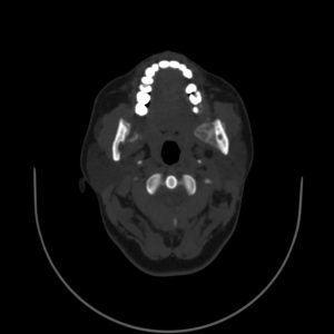
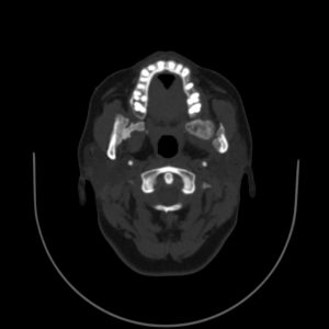
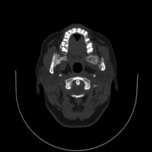
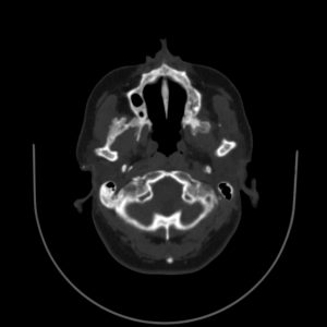
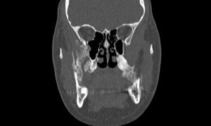
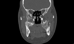
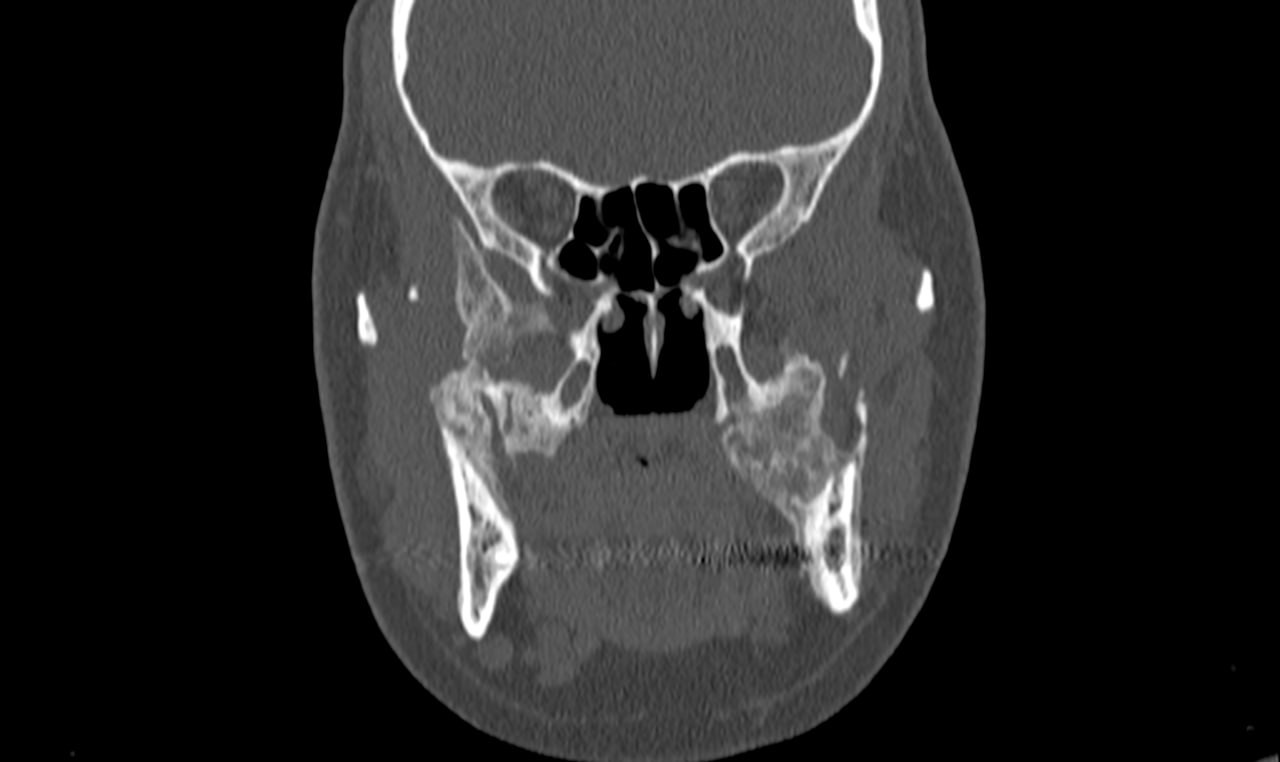
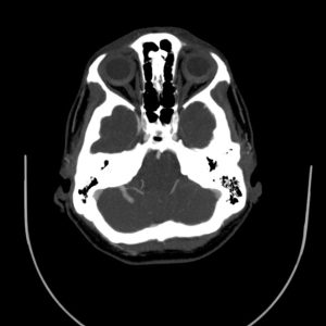
.Heterotopic Multicentric ossification of soft tissue is noted in patient’s face. This ossification has involved subtemporalis fossa at both sides
Right masseter muscle, right lateral pterygoid, left medial and lateral pterygoid and left masseter muscle are involved in this multicentric heterotopic ossifcation. Also, involvement of inferior part of right temporalis muscle is detected
.Some heterotopic ossifications are also present adjacent to condyle of left mandible, which can lead to malocclusion
.Most of These ossified soft tissues show ground glass density
These findings can be in favor of a heterotopic ossification process such as myositis ossificans traumatica of masticatory muscles . Correlation with previous imagings and clinical history is recommended
:Diagnosis: (with pathology confirmation)
Myositis ossificans traumatica
:Myositis ossificans traumatica
Male patients have been considered as the main group at risk of developing MOT of the masticatory muscles with a male/female ratio of 2.4/1
MOT has been frequently related to traumas (e. g. fracture, blow) a possible explanation could be: males might have experienced traumas
more often than females
.prevalence for female patients of MOT of the masticatory musculature in context of dental treatment with a 1.5/1 ratio
In most cases of MOT of the masticatory muscles the masseter muscle is the most affected one. However, this is not true for those
. cases of MOT occurring after dental treatment. Of these cases, 66% involved the medial pterygoid muscle
:References
https://head-face-med.biomedcentral.com/articles/10.1186/s13005-018-0180-6
/https://www.ncbi.nlm.nih.gov/pmc/articles/PMC4196299
